What are Phantoms?

Imaging phantoms or simply "phantoms" are specially designed objects that are scanned or imaged in the field of medical imaging to evaluate, analyze, and tune the performance of various imaging devices. These objects are more readily available and provide more consistent results than the use of a living subject or cadaver, and likewise avoid subjecting a living subject to direct risk.

Phantoms were originally employed for use in 2D X-ray-based imaging techniques, such as radiography or fluoroscopy, though more recently phantoms with desired imaging characteristics have been developed for 3D techniques, such as MRI, CT, Ultrasound, PET, and other imaging methods or modalities.

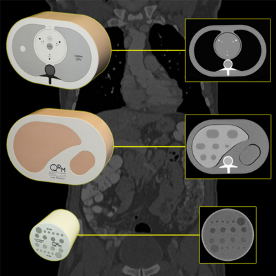
QRM-Cardio-Thorax, Liver and Low-Contrast Insert. Phantoms for measuring coronary artery calzifications, liver lesions and low contrast capability of CT scan protocols.

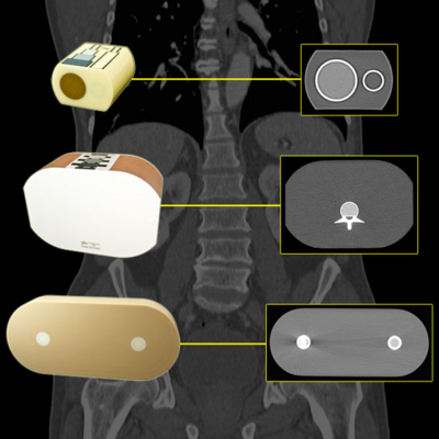
Phantoms for CT and DXA to measure the BMD (Bone Mineral Density)

A phantom used to evaluate an imaging device should response in a similar manner to how human tissues and organs would act in that specific imaging modality. For instance, phantoms made for 2D radiography may hold various quantities of X-ray contrast agents with similar X-ray absorbing properties to normal tissue to tune the contrast of the imaging device or modulate the patient's exposure to radiation.

In such a case, the radiography phantom would not necessarily need to have similar textures and mechanical properties since these are not relevant in X-ray imaging modalities. However, in the case of ultrasonography, a phantom with similar rheological and ultrasound scattering properties to real tissue would be essential, but X-ray absorbing properties would not be needed.

Article from Wikipedia - The Free Encyclopedia!
--> Imaging phantom

Anthropomorphic phantoms are physical test objects simulating anatomical structures of the body, which makes them easier and more convenient to use. Some phantoms provide equivalent properties as the real human body does, e.g., organs, such as lung, liver, fat, bone. This is an advantage in X-ray imaging due to dose issues – medical devices can be thus tested without using human volunteers.

Physical parameters of such devices can be tested as well in a more physical or technical way, for example the spatial resolution of a system can be tested with a wire in a cylindrical phantom to create a MTF, or with a high resolution bar pattern as well for visible testing.

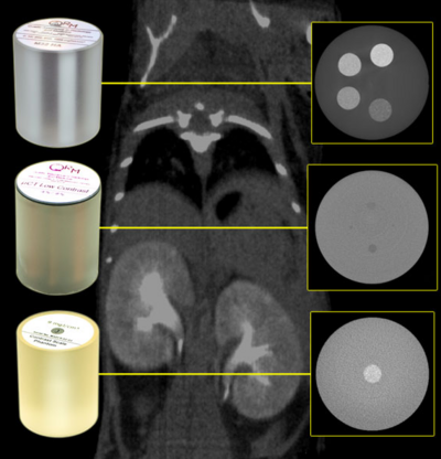
Phantoms for Micro-CT systems to calibrate bone densities and other materials as iodine, and to test low contrast capabilities of the scanner.

Diese Seite existiert auch in Ihrer Sprache.
This page also exists in your language.
Sprache wechseln Change Language